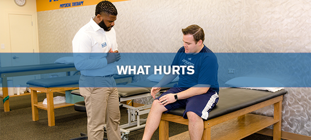
What Hurts?
 Questions About Your Injury Or Pain?
Athletico has created an injury hotline available to anyone who sustains an injury or has questions about pain, strains or injuries. A certified and licensed athletic trainer (AT) will answer your questions and direct you appropriately for care.
Call Athletico’s Injury Hotline at 1-877-ATHLETICO (877-284-5384).
Click here to find a Location near you.
Select a category below to learn more about injuries.
- Head & Neck Injuries
- Shoulder Injuries
- Elbow/Forearm Injuries
- Low Back Pain
- Wrist/Hand Injuries
- Hip Injuries
- Knee Injuries
- Foot Injuries
- Lower Leg Injuries
- Ankle Injuries
Head & Neck Injuries
Cervicogenic Headache:
This condition describes an individual who is experiencing headaches due to dysfunction within the upper cervical spine (neck). Patients typically report moderate to severe unilateral (one sided) pain symptoms provoked with sustained neck postures, stress, and neck movement that increases in intensity as the day progresses. Pain usually starts in the suboccipital region (base of the neck) and radiates into the front of the head and behind the eyes. Patients may experience these headaches anywhere from daily to 2-3 times per week with symptom duration ranging from 3-24 hours per day. Additional systemic symptoms that may be present include nausea, vomiting, phonophobia (sensitivity to sound), photophobia (sensitivity to light) and blurred vision. Significant evidence has been published demonstrating the utilization of specific physical therapy interventions to the neck (therapeutic exercise and manual therapy techniques) on the reduction of cervicogenic headache frequency, intensity, disability, and medication intake.
Cervical Radiculopathy:
This condition describes an individual who is experiencing upper extremity weakness, numbness, pain, and/or muscular atrophy associated with disease to the nerve roots of the cervical spine (neck). The patient experiencing cervical radiculopathy will generally describe sharp, lacerating pain symptoms in the affected arm with no known cause. The most common direct pathological sources of radiculopathy include a cervical disc herniation and osteophyte formation (bone spurs) within the cervical spine. The incidence of this condition is most frequently seen in patients in the fifth decade of life (2 %). This rate is three times higher than in any other age group.
Acceleration Injury (Whiplash):
The basic mechanism of a whiplash injury involves acceleration of the head and cervical spine (neck) relative to the rest of the body. A common occurrence resulting in an acceleration injury involves the collision of two motor vehicles; however, whiplash may also occur as a direct result of participation within high-velocity, contact sports such as football. The classic mechanism of injury, however, is a passenger within a motor vehicle struck from behind. Most patients report significant neck pain, stiffness, and muscle spasm occasionally accompanied with headaches. Symptom onset is typically experienced within two days following the initial injury.
Thoracic Outlet Syndrome:
This condition consists of compression of the neurovascular structures (nerves and arteries) of the neck and shoulder as they travel through cervicoaxillary region. Symptoms that accompany this condition can be highly variable with the presentation depending on where the neurovascular compression is occurring. Patients often report numbness and pain from the neck and shoulder down along the inside portion of the forearm and hand. Additional symptoms that may be present include intrinsic hand muscle weakness and atrophy, a diminished pulse, and decreased blood pressure in comparison to the unaffected side. Common causes of this condition include congenital abnormalities (e.g. presence of a cervical rib) and trauma to either the clavicle or the first rib.
Athletico offers free assessments. LEARN MORE.
Click to Request an Appointment Today
Shoulder Injuries
Rotator Cuff Tendonitis:
This condition is often associated with repetitive, abnormal stress to the tendons of the rotator cuff (four small muscles that surround and steer shoulder movement) resulting in inflammation and pain. Resultant cuff tendonitis may cause sharp, acute pain in the shoulder or upper arm aggravated after periods of activity such as overhead throwing or lifting. Pain may also be experienced when dressing, grooming, sleeping on the affected shoulder, reaching high over head, or behind the back. Functional weakness is usually present with lifting during everyday activities (especially between waist and shoulder height). If the condition is left untreated, the tendonitis may progress to a partial thickness tear of the rotator cuff, often requiring surgery. Physical therapy can be beneficial to regain lost shoulder motion and functional strength while decreasing pain and facilitating the healing process to the injured tissues.
Anterior Shoulder Dislocation:
This injury typically occurs as a direct result of trauma to the ligaments and capsular tissues that surround the ball and socket (glenohumeral joint) of the shoulder. Some common mechanisms of injury include being hit behind the arm while the shoulder is positioned in an overhead throwing motion and falling onto an outstretched arm. This condition contributes to a sense of instability in the shoulder combined with an inability to perform certain daily activities and sports. Those who experience a shoulder dislocation are typically evaluated by a physician for reduction and to rule out fracture or cartilage (glenoid labrum) damage. Physical therapy is often ordered to help restore shoulder motion and strengthen the muscles that cross the shoulder to prevent recurrence of dislocation.
SLAP (Superior Labrum Anterior and Posterior) Lesion:
This condition involves injury to the superior (top) portion of the labrum of the shoulder joint. The labrum is a cartilaginous ring that serves to deepen the socket of the joint providing both stability and a site for muscular attachment for the biceps brachii. Common causes of a SLAP lesion include falling onto an outstretched hand, overhead lifting, and overhead throwing. This injury can be difficult to identify clinically; however, common patient reports include instability within the shoulder causing a vague ache. In addition, some patients may report catching, popping, or clicking within the joint during functional activities.
Rotator Cuff Impingement:
This condition involves a progressive, mechanical impingement of the rotator cuff tendons beneath the bony architecture (coracoacromial arch) of the shoulder joint. The resultant impingement of the cuff tendons results in significant shoulder pain increased with the performance of overhead and functional activities. Common causes of cuff impingement include bony abnormalities and rotator cuff tendon thickening. Conservative treatment is typically geared towards decreasing the initial pain and inflammation, restoring pain free range of motion within the shoulder, and rebuilding functional strength to the rotator cuff and scapular musculature.
Adhesive Capsulitis (Frozen Shoulder):
This condition involves stiffening (freezing) and inflammation of the soft tissues (joint capsule and ligaments) that surround the shoulder joint. The stiffening of these structures creates severe loss of functional shoulder movement, pain surrounding the joint, and an inability to sleep on the affected side. The time for complete resolution of shoulder range of motion can vary between 12 to 36 months. The incidence for this condition is approximately 2% within the general population and from 10-35% within the diabetic patient population. Other common factors related to an increase in the prevalence of this condition include cervical spine (neck) disorders, hypothyroidism, and prolonged post-surgical or post-traumatic immobilization of the shoulder.
Rotator Cuff Tear (Partial Thickness and Full Thickness):
This condition involves complete (full thickness) or incomplete (partial thickness) disruption of the tendons of the rotator cuff muscle group. Common causes of injury include direct trauma to the shoulder, repetitive overhead lifting, and participation in sports that require overhead throwing. In addition to these causes, some patients experience a cuff tear simply as a direct result of a degenerative process with no specific trauma or activity associated with the injury. A common presentation for a patient with a rotator cuff tear includes an individual 40 years of age or older with reports of constant, lateral shoulder pain affecting the ability to sleep accompanied with functional weakness limiting his or her ability to lift the arm against gravity.
Acromioclavicular Joint Injuries:
The acromioclavicular (AC) joint (the connection between the collar bone and the shoulder blade) is commonly injured as a result of either a direct force to the tip of the shoulder or through an indirect force sustained during a fall on an outstretched hand. This resultant force results in disruption to the capsule and ligaments that supports the bony architecture of the AC joint. The patient with an acute AC joint injury will typically cradle the involved arm against the body with the uninvolved hand for support. This posture helps to decrease the pull of the weight of the arm against the ligamentous and capsular tissues that have been disrupted.
Athletico offers free assessments. LEARN MORE.
Click to Request an Appointment Today
Read articles on the shoulder on the Athletico Blog.
Elbow/Forearm Injuries
Tennis Elbow:
Also known as lateral epicondylitis. Tennis elbow stems from overuse, improper muscle strength, and repetitive movement of the wrist or elbow where the tendons at the elbow become stressed due to poor mechanics (i.e. typing, racquetball, tennis, golf). Localized pain at the lateral (outside) elbow is present with wrist and elbow movement. Pain can become so intense that lifting a glass of water may be a chore! Tennis elbow can be difficult to relieve if mechanics and flexibility/strength issues are not addressed. Ice massage and anti-inflammatories may help with the acute pain, but therapy may be required to address proper work and leisure ergonomics as well as muscle imbalances. Thermal or electrical modalities may be used to decrease inflammation and promote tissue healing. Splinting of the wrist may also be utilized to rest the muscles around the elbow during this time of healing. Many sporting goods stores carry straps that are placed below the elbow to help reduce pain. While these straps may be beneficial for some, they should not be considered as a cure-all, and improper use may even worsen symptoms. Our professional staff will also be able to analyze your golf swing or racquet stroke to prevent further injury.
Golfer’s Elbow:
Also known as medial epicondylitis. Similar to tennis elbow with associated pain and decreased movement, but golfer’s elbow occurs on the inside of the elbow. Golfer’s elbow presents similar signs and symptoms as tennis elbow and is also difficult to heal if not handled properly. Therapeutic management of golfer’s elbow is very similar to that of tennis elbow. Splinting may be used to decrease strain on the muscles, and the use of anti-inflammatories will help with tissue swelling and pain. Therapy focuses on restoration of muscle balances (flexibility and strengthening), education on causative factors and prevention, and thermal and electrical modalities to decrease inflammation and facilitate healing. Our professional staff will also be able to analyze your golf swing or racquet stroke to prevent further injury.
Pronator Syndrome:
This condition involves the compression of the median nerve in the forearm. The median nerve passes into the forearm down the front of the elbow and passes under ligaments and into muscles. Compression of this nerve in the forearm generally occurs as it enters between two heads of a muscle—the pronator teres (thus the name “pronator” syndrome). The pronator teres muscle turns the palm of the hand down. Patients with this condition usually complain of an aching pain in the forearm, increased pain with gripping while the forearm is pronated (palm down), decreased strength, and forearm fatigue. Conservative management is generally the first course of treatment. Occupational therapy may be prescribed for splint fabrication to limit forearm rotation, nerve gliding exercises, stretches to maintain flexibility, and activity and job modification to restrict repetitive forearm rotation. If the condition is unresponsive to conservative treatment, surgical decompression of the nerve may be considered.
Cubital Tunnel Syndrome:
This condition involves the ulnar nerve as it travels down the inside of the arm behind the elbow. This nerve lies in a groove on the inside of the elbow. If you’ve ever hit your “funny bone” then you’ve experienced the symptoms of Cubital Tunnel Syndrome—elbow pain and numbness/tingling in the ring and small fingers. If left untreated, it can progress to significant hand weakness and a “claw” deformity of the ring and small fingers. It is exacerbated by repetitive or static bending of the elbow, arthritis, or trauma to the elbow. Treatment for Cubital Tunnel Syndrome includes use of anti-inflammatories to reduce swelling, splinting of the elbow at night to prevent bending of the elbow and stretching of the nerve, and use of an elbow pad during the day to protect the nerve. You may be referred to an occupational therapist for splint fabrication, education on proper body mechanics and workstation set-up, nerve gliding exercises, and exercises to maintain range of motion and strength.
Fractures:
Fractures may be caused by falling on an outstretched arm or by direct trauma to the elbow. The elbow has three joints that are surrounded by ligaments. Because of its complex structure, improper alignment of the bones and any associated ligament damage can significantly reduce elbow motion, stability, and function. It is very important to seek medical attention if an elbow fracture is suspected.
Athletico offers free assessments. LEARN MORE.
Click to Request an Appointment Today
Low Back Pain
The spine is the most dynamic structure in our body as it allows for optimal stability and movement and maintains our posture. This highly integrated and dynamic structure is involved in all movement and is undergoing constant stress 24/7, so it should not be surprising to us when it starts to show wear and tear. Contributing factors to low back pain include poor body mechanics and work ergonomics, decreased strength, and muscular/structural imbalances. As the low back is subject to repetitive stresses of daily life and occasional injury, it may respond by showing signs and symptoms of wear and tear. These symptoms may manifest themselves as pain located around the waistline and/or lower extremities, and possibly, numbness and tingling to the lower extremities. If any of these symptoms persist or worsen, seek immediate medical attention.
Spondylolisthesis:
Spondylolisthesis is a forward slipping of a vertebrae over another. The individual may or may not present with pain and is usually the result of repetitive stress to the spine. A program emphasizing proper body mechanics, aerobic conditioning, lumbar stabilization, core strengthening, and patient education is key to eliminating and preventing low back pain. Individuals with low back pain are encouraged to take a proactive role in their recovery in conjunction with the guidance and supervision of your physician, physical therapist, and athletic trainer.
Low Back Strain:
Muscle strains may occur as a result of poor body mechanics with daily activities/work tasks or unexpected movement (i.e. lifting heavy objects, slipping on ice, or motor vehicle/sports accident). All of these can cause the muscles to contract beyond their normal limits placing stress on the spine. Ice and anti-inflammatories will help reduce pain and swelling. Consult a physician, physical therapist, or an athletic trainer to determine severity of injury and if exercise may be beneficial.
Herniated Disc:
Discs are located between the vertebrae of the spine to help minimize shock and help optimize movement. As we age, the discs lose their elasticity and may tear or bulge onto the spinal nerves. This can produce extreme muscle spasms and pain to low back and legs and/or numbness and tingling to legs and toes.
Athletico offers free assessments. LEARN MORE.
Click to Request an Appointment Today
Wrist/Hand Injuries
De Quervain’s Syndrome:
This condition involves inflammation of the tendons of the thumb. Pain is very noticeable in the wrist and thumb during general thumb use and during gripping and pinching activities. Conservative management usually consists of splinting the wrist and thumb, along with the use of anti-inflammatories (oral and/or injection). Occupational therapy may also be introduced to restore flexibility to the wrist and thumb, evaluate causative factors, educate on the prevention of symptoms, and strengthen the wrist and thumb to regain function. Therapy may also be prescribed to use thermal or electrical modalities to control inflammation and pain.
Fractures to Hand or Wrist:
A common mechanism of injury is falling on an outstretched arm with the wrist hyper-extended. Proper alignment of the bone(s) is essential for normal healing and restoration of motion. In addition, because of important vessels and nerves surrounding these structures, it is very important to follow-up with an orthopedic surgeon or a hand specialist. Treatment generally consists of casting or surgery to stabilize the fracture, followed by therapy to regain range of motion of the joints.
Tendonitis:
Tendonitis, simply put, is inflammation of the tendon. A tendon is what connects muscles to bone, and it typically crosses a joint. Overuse of the joint or muscle causes inflammation of the tendon. Tendonitis is very common in the wrist and hand. Tendonitis of specific tendon(s) can have different names (i.e. DeQuervain’s tenosynovitis, Intersection syndrome, finger tendonitis), but treatment is generally the same.
Tendonitis is generally treated with anti-inflammatories to reduce pain and swelling as well as by immobilizing the joints the tendon crosses. Occupational therapy may be prescribed to use thermal or electrical modalities to decrease pain and inflammation, for custom splint fabrication, to learn exercises and stretches to restore muscle and tendon flexibility, and to strengthen the wrist and hand to resume normal use. Your workstation and daily activities may need to be modified to prevent further injury and overuse.
Carpal Tunnel Syndrome:
The carpal tunnel is a narrow passageway in your wrist that allows nine tendons in the fingers and thumb, as well as the median nerve, to travel into the hand. Pressure inside the carpal tunnel may be increased by repetitive wrist motions, gripping, or sustained wrist and finger positions. This increased pressure on the nerve may cause wrist pain, numbness and tingling in the thumb and first two fingers, and eventual hand weakness.
Carpal Tunnel Syndrome may be managed with anti-inflammatories and with splinting to immobilize the wrist and decrease pressure in the carpal canal. A patient may be referred to an occupational therapist for splinting, nerve and tendon exercises, thermal or electrical modalities to decrease inflammation, and education on prevention of symptoms and activity modification. Our professional staff may also visit your worksite to adjust your workstation and fully optimize good technique to avoid future injury or recurrence. If conservative management is unsuccessful, surgery may be required to decompress the nerve.
Arthritis:
There are many forms of arthritis with most forms being categorized as either Osteoarthritis or Rheumatoid arthritis. Osteoarthritis generally occurs from “wear and tear” on the joints, while Rheumatoid arthritis is actually an autoimmune disorder that attacks the lining of the joints. Both forms of arthritis frequently occur in the wrist and hand. In addition to medical management, occupational therapy may be prescribed. Therapy goals are to decrease joint inflammation, improve joint range of motion, and provide education on joint protection techniques as well as to provide equipment to relieve strain on the affected joints during daily activities. Therapists may also fabricate rigid splints to rest and immobilize joints during a “flare-up” and recommend a variety of soft splints that support joints during hand use.
Trigger Finger:
Trigger finger, also known as stenosing tenosynovitis, can occur in any of the fingers or thumb. It is caused by the swelling of one of the tendons that bend the finger or thumb. This tendon inflammation causes the finger to catch in a bent position. Straightening of the finger will then cause it to snap. Trigger finger can be associated with chronic inflammation (i.e. rheumatoid arthritis), overuse of the hand, or from using tools with hard or sharp edges. Conservative management may consist of anti-inflammatories or cortisone injections. Occupational therapy may be prescribed for splinting of the hand in order to rest the tendon and prevent triggering, for use of modalities to decrease inflammation, to provide exercises to maintain joint motion, and to provide ergonomic assessment and education.
Mallet Finger:
Mallet finger is an injury to the fingertip. It commonly occurs when the tip of the finger is hit—usually while playing sports such as baseball and basketball. With this injury, the tendon that straightens the tip of the finger is disrupted, and the finger ends up in a bent position. Mallet finger can also be associated with a fracture of the fingertip. Your finger will be splinted or pinned in a straight position until the tendon heals (usually around 6 weeks). The tip of the finger is NOT allowed to bend during this time. You may need to see a specialist if there is a fracture or if the finger does not heal properly.
Tendon/Ligament Injuries to Fingers:
These types of injuries usually occur with direct contact to the fingers (“jammed finger”) or forceful gripping of an object that is moving. Pain may occur with movement, or in some cases, finger movement may not occur at all if a tendon is ruptured. Proper medical attention is necessary to avoid permanent deformity to the finger involved. Immobilization is usually done as required by the physician to allow proper healing of the damaged tissues. Once the splint is removed, occupational therapy will help restore proper motion to the fingers and facilitate the return to full function.
Guyon Canal Syndrome:
This condition involves the ulnar nerve and artery as they pass into the hand at the wrist (on the small-finger side of the hand). It is also known as “handlebar palsy”—named for pressure on the ulnar nerve in the hand from the handlebars of a bicycle during long-distance cycling. Pressure on this nerve causes numbness and tingling in the ring and small finger, pain on the small-finger side of the hand, and eventual hand weakness.
Treatment generally consists of conservative management with the use of anti-inflammatories and/or therapy. Therapy may include splinting of the wrist until the irritation of the nerve subsides, use of modalities to decrease inflammation, activity and tool modification to reduce pressure on the nerve, and strengthening of weak muscles.
Athletico offers free assessments. LEARN MORE.
Click to Request an Appointment Today
Hip Injuries
Hip Pointer:
A bruise caused by direct contact to the front hip bone. This injury can produce extreme pain and limit normal motion at the hip and trunk. Because of close proximity of internal organs, follow-up with a family physician is advised to rule out potential fracture or internal injury. Range of motion exercises may begin to help minimize pain and swelling if all other injuries are ruled out. Ice and anti-inflammatories will also help reduce the effects of the injury.
Piriformis Syndrome:
This condition refers to irritation of the piriformis muscle which lies underneath the gluteus muscle, or buttock. Because the sciatic nerve passes underneath or through the piriformis muscle, burning or numbness/tingling may occur due to nerve irritation. Pain may start in the buttock and radiate down the affected leg. It is important to seek proper medical attention to rule out referred pain from the spine. In most cases, piriformis syndrome may be alleviated through anti-inflammatories, and lower extremity flexibility program, if spine problems are ruled out.
Iliotibial Band Syndrome:
Inflammation of the thick, fibrous tissue that runs from the top of the hip to just below the knee. This injury commonly occurs in runners and can be very debilitating. Formal rehabilitation may be required to reduce pain and inflammation and restore proper muscle balances throughout the pelvic region. Once the pain diminishes, a thorough running analysis may be completed to prevent recurrence of the injury.
Groin:
An injury to the inner thigh caused by running, jumping, twisting, kicking. Signs and symptoms may range from mild tenderness over the muscle involved to the inability to contract the muscle and walk. There will be localized pain and swelling and in severe cases, the person may have bruising to the inner thigh, indicating bleeding to the injured muscle. Depending upon the severity, treatment may include ice, compression wrap, crutches (if unable to walk). Rehabilitation is usually slow and controlled to minimize re-injury.
Quad:
Injury to the front of the thigh caused by a forceful contraction of the quad muscle when the hip is bent and the leg is straight (as in a kicking movement). Signs and symptoms may range from mild tenderness to the touch to the inability to walk without pain. Bruising may be present with associated swelling. Treatment includes ice, anti-inflammatories, crutches (if unable to walk). In severe cases, rehabilitation may be warranted to regain proper mobility.
Strains:
Injuries to the muscles and their tendons due to a forceful contraction of the muscle involved.
Athletico offers free assessments. LEARN MORE.
Click to Request an Appointment Today
Knee Injuries
Osteoarthritis (OA):
General degeneration of the knee joint that stems from wear and tear. This is accompanied by gradual increase in pain with activities (walking, stairs, prolonged sitting or standing). Physical therapy will help alleviate the pain by focusing on proper strengthening.
ACL Injury:
The ACL injury is the most common injury to the knee. This ligament prevents the lower leg from moving forward on the upper leg. The mechanism of injury is from a twisting motion when the foot is firmly planted. The degree of severity ranges from a mild stretch of the fibers (Grade I) to complete rupture of the ligament (Grade III). The individual may feel or hear a “pop” with associated swelling. The individual may also report a feeling of “giving out” to the knee, limiting the function of the knee. An orthopedic specialist will be able to help diagnose and recommend treatment options for the individual.
MCL Injury:
This ligament lies along the inside of the knee and can be damaged by direct trauma from the outside of the knee towards the inside while the foot is planted. The individual may experience pain while walking, climbing stairs, and prolonged sitting. Treatment may include ice, anti-inflammatories, and short-term immobilization to allow the fibers to heal. Rehabilitation may be required to establish range of motion and strength following this injury.
Meniscus Injuries:
Commonly called torn cartilage, this is an injury to one of the two circular pads between the upper and lower legs. They function to decrease shock to the knees and distribute weight bearing forces through the legs. The mechanism of injury is a compression force associated with a twisting motion. A “pop” may be heard, but there is usually increased pain along the joint line of the knee. Signs and symptoms include pain, swelling, knee “locking up” or the feeling that the knee is stuck, and difficulty with walking and stairs. An orthopedic consultation is suggested to rule out trauma to other structures. If the tear is small enough, the meniscus may heal on its own; however, if pain and decreased function persist, arthroscopic surgery may be required to repair the meniscus.
Sprains:
Injuries to the ligaments in a joint. The knee has four main stabilizing ligaments, any of which can cause problems if torn.
Patellofemoral Pain Syndrome:
Categorized as general knee pain that manifests itself near or around the kneecap (patella). This can be caused by an acute injury, muscle imbalances, walking abnormalities, or misalignment of the kneecaps. One or a combination of all of the above can lead to PFPS, causing pain with walking, stairs, squatting, prolonged sitting. Physical therapy can address muscle imbalances and acute symptoms, as well as recommend proper footwear to maximize proper walking mechanics.
Athletico offers free assessments. LEARN MORE.
Click to Request an Appointment Today
Foot Injuries
Plantar Fasciitis:
Catchall phrase used to describe pain along the inside arch and heel. Increased pain occurs upon the first few steps in the morning or after prolonged sitting. Plantar fasciitis can occur in individuals with poor foot biomechanics (excessive foot pronation), decreased flexibility and strength in the surrounding musculature. Ice massage and rolling the foot over a rolling pin may help reduce pain and irritation. Proper shoe wear and walking analysis should be addressed as well.
Turf Toe:
Repetitive overuse or trauma to the Big Toe, causing the toe to hyperextend. This condition can be very painful, especially with walking or recreational activities involving stop and go movements. Orthotics may help decrease the pain limiting the toe’s movement. Ice and anti-inflammatories will also help reduce pain and swelling to the joint.
Morton’s Neuroma:
A mass of tissue that surrounds the nerve sheath of the nerves that innervate the toes. It commonly occurs between the third and fourth toes and can cause burning, numbness, and tingling to the toes. Proper shoe wear is a must to avoid compressing the toes. Ice and anti-inflammatories will also help decrease the inflammation surrounding the nerve.
Athletico offers free assessments. LEARN MORE.
Click to Request an Appointment Today
Lower Leg Injuries
Shin Splints:
Overuse musculotendinous injury that can be caused by abnormal biomechanics, poor conditioning, and improper training. Pain can manifest in two locations: anterior or posterior.
Anterior shin splints:
Pain is on the outside of the lower leg and is associated with those runners who have a loud heel strike when running.
Posterior shin splints:
Pain is on the inside of the lower leg toward the ankle. In both instances, weak muscles in the lower leg accompanied by improper footwear and running mechanics may exacerbate the symptoms and possibly lead to stress fractures. Physical therapy is needed to focus on pain reduction, lower extremity flexibility and strengthening, proper running mechanics, and proper footwear.
If the above factors go unchecked, stress fractures may occur. They are usually caused by fatigue and breakdown of the surrounding musculature, subsequently placing greater stress on the bones. Immobilization is required to allow the bone to heal. Surprisingly, stress fractures do not appear on X-rays until after the fracture site starts to heal.
Achilles’ Tendonitis:
Inflammation of the Achilles’ tendon caused by overuse injuries (running, jumping), as well as decreased calf strength and flexibility. Tenderness and swelling are present over the tendon along with pain with walking, stairs, and running. Treatment includes ice, anti-inflammatories, flexibility training, and lower extremity strengthening. Foot biomechanics and proper footwear should be addressed as well.
Athletico offers free assessments. LEARN MORE.
Click to Request an Appointment Today
Ankle Injuries
Lateral Ankle Inversion Sprain:
This injury is typically found in athletics when an individual “rolls” their ankle. Often times, this injury is characterized by swelling at the outside ankle bone (lateral malleolus) with possible bruising if the injury is severe enough. An ankle sprain results in damage to the ligaments of the outer ankle, although a small fracture can occur at the outer ankle bone. Initial management of this injury should include rest, ice with compression, and elevation of the leg to decrease swelling.
Achilles Tendonitis:
The Achilles tendon is the large tendon found at the back side of the foot and ankle. This tendon attaches the calf muscles to the ankle and allows the ankle to be pointed in a downward position and propel us when we walk. This tendon injury is characterized by pain with walking and swelling and tenderness at the tendon. The tendon will typically feel worse with walking and running.
Peroneal Tendonitis:
The peroneal tendons are the tendons at the outside of the ankle. In some instances, these tendons can become injured resulting in swelling and pain at the outside of the ankle, slightly underneath the outer ankle bone. These tendons can also be injured following a lateral ankle sprain when the peroneal muscle is forced to contract quickly to prevent the ankle from rolling inward too far. Ice and compression is helpful in managing this injury acutely but if pain persists, seeking medical attention may be helpful in resolving the injury completely.
Morton’s Neuroma:
A neuroma is a small tumor like cyst that forms around the sensory nerve that is found between the third and fourth toes typically. The symptoms of a Morton’s neuroma are tingling and numbness occurring between the toes made worse with running or walking. A medical consult should be sought if symptoms persist.
Posterior Tibialis Tendonitis:
The posterior tibialis muscle is found at the inner aspect of the lower leg with the tendon extending down the leg and along the inner aspect of the foot. The function of this muscle and tendon is to support the arch of the foot. This tendon can become injured with running and also if the foot pronates or collapses too much. In this instance, the muscle and tendon become overworked resulting in swelling and irritation of the tendon. Often, this injury requires a biomechanical assessment by a medical professional to resolve aggravating factors and resolve the injury. Initial management can include ice and rest.
Plantar Fasciitis:
The plantar fascia is a thick band of tissue along the arch and the bottom of the foot that is needed to support the arch. This band of fascia attaches the underside of the heel. This diagnosis is usually used to describe pain that occurs at the inside arch and the heel. Typically, this is characterized by pain occurring at the heel with the first step in the morning and made worse with prolonged walking and running. Ice massage at the area of the pain can be helpful and analysis of biomechanical factors contributing to the injury is usually needed to completely resolve this condition.
Stress fractures:
A stress fracture can occur in any bone; however, one area common for injury is at the 5th metatarsal or the lateral bone of the foot. Usually the area of the fracture is point tender and feels worse with running and sometimes with walking. A stress fracture is typically diagnosed with the use of a bone scan or an MRI.
Athletico offers free assessments. LEARN MORE.
Click to Request an Appointment Today